 A gynaecology ultrasound or pelvic ‘scan ‘ is a detailed imaging assessment of the female pelvis. This includes imaging of the vagina, bladder, urethra, uterus, endometrium and ovaries.
Abdominal and Vaginal Ultrasound
The structures in the female pelvis can be viewed with an ultrasound probe on the abdomen, on the perineum or in the vagina. A typical examination involves using both approaches to obtain the highest quality imaging.
An ultrasound probe placed in the vagina usually produces the clearest imaging of the pelvic anatomy. The closer an ultrasound probe is to a structure, the higher quality ultrasound picture can be produced.
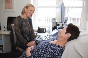
The doctor or sonographer performing the ultrasound will discuss the reasons for your referral to us prior to commencement of the examination.
The type of ultrasound most appropriate to answer your health concerns will be discussed and options for the individual women offered.
Abdominal imaging
There are many situations for an individual female where a vaginal ultrasound is not able to be performed or completed. The specialist will be happy to discuss your questions regarding this at the beginning of the consult.
If you are being referred for an abdominal ultrasound of the pelvis please attend with a full bladder. The bladder provides an ultrasound ‘ window ‘ to allow high quality imaging of the pelvic structures.
Vaginal imaging
The ultrasound probe is a narrow gel covered instrument that is gently placed several centimetres into the vagina. The probe is placed close to the cervix and is carefully moved to obtain clear images of the pelvic anatomy. Most women would experience minimal discomfort. The ultrasound takes a little longer than a pap smear examination, but would be intended to be a more comfortable procedure.
Indications for gynaecology ultrasound
- Pelvic pain
- Abnormal vaginal bleeding
- Heavy menstrual bleeding
- Irregular cycles
- Bleeding between menstruation
- No menstrual bleeding
- Screening for ovarian cancer or cysts
- Infertility
- Past or Family history of gynaecological conditions
- Fibroid Surveillance
Adolescent and Paediatric Gynaecology Imaging
The imaging team at Specialist Imaging for Women work with the medical teams at the Royal Children’s Hospital Melbourne, Mercy Hospital for Women and Royal Womens Hospital to provide sub specialist imaging for young women. This is typically an abdominal Ultrasound where the pelvic organs are imaged with a full urinary bladder. The specialist is available at the time of the ultrasound to discuss the report findings.
Deep Endometriosis Imaging
Endometriosis is thought to affect around 10 % of women. This incidence increases to 30 % in women with infertility. The symptoms of endometriosis include pelvic pain, pain with bowel movements, pain with urination, pain with sex and painful periods. Early diagnosis has been shown to benefit women with prompt management strategies to control symptoms.
Whilst laparoscopy remains the gold standard investigation for diagnosis , there have been significant improvements in medical imaging in the diagnosis of endometriosis.
Our imaging team work in conjunction with the tertiary endometriosis units at the Mercy Hospital for women and Royal Womens Hospital to provide information to surgical teams about diagnosis and determining the extent of disease.
Dr Kate Stone is a member of SAFE ( www.safe-endo.com.au ) and The Pelvic Pain Foundation of Australia ( www.pelvicpain.org.au ) and is actively involved in research in endometriosis.
The ultrasound assessment for endometriosis involves a normal transvaginal scan as described above. As endometriosis can infiltrate the bowel or rectal wall this will also be assessed via the transvaginal imaging at the time of the scan. The images obtained of the rectal wall are generally improved if the bowel is empty.
In women with a suspicion or prior diagnosis of deep infiltrating endometriosis affecting the rectum, your referring doctor may request that you have a fleet enema prior to your ultrasound. If you do not have a past history of significant deep endometriosis or no bowel symptoms you most likely do not need to take the bowel preparation.
No oral laxative is required.
If you have any queries as to whether you need to use a fleet enema ( bowel preparation ) prior to your ultrasound please contact our staff or referring doctor. If you have been asked to use a fleet enema prior to your ultrasound, you can find our information sheet here