 12 Week Ultrasound
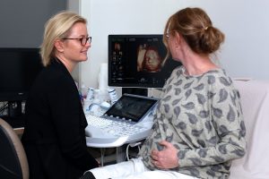
An Ultrasound at 12-13 weeks allows the early fetal anatomy to be seen very clearly. As well as assessing fetal number, heart rate, placental site and a check of maternal ovaries, a detailed scan of the early fetal structures is performed.
It is possible to see the 4 chambers in the heart, the heart valves and the major blood vessels arising from the heart. Early fetal brain is assessed as well as spine, arms, legs and even hands. The improvement in ultrasound technology has enabled fetal structures that were only seen at the 20 week scan to be examined much earlier.
A measurement is taken of the nuchal translucency, which is the normal collection of fluid present in all fetuses at this gestational age. This measurement can be used to assess the risk for T21 (Downs Syndrome) for the first trimester combined serum screen and as a method of assessing fetal wellbeing as it may be thickened for a variety of reasons.
This examination can be performed with a transabdominal scan, a transvaginal scan or a combination both depending on how the best imaging is obtained.
Most major structural abnormalities can be detected at this gestation with good imaging. This scan does not replace the 20 week scan which allows even more detailed assessment of the fetus when it is bigger and more mature.
The scan takes approximately 30 minutes and there is no preparation.
Note: Additional time is required for a multiple pregnancy. Please advise our staff when booking if you are carrying twins or triplets.
20 Week Ultrasound
A morphology or anatomy scan is performed at 20-21 weeks gestations. At this stage the fetus is large enough to examine most of the developing anatomy in detail. The examination looks at the physical structure of the fetus rather than function. The heart, brain, spine, limbs and face are assessed as well as kidneys and bladder. It is possible to look at many of the internal abdominal organs as well. A large number of measurements are taken, some of these relate to the size of the fetus and some relate to structures being normally developed. Patients may request to find out the gender of the fetus if they wish.
The amniotic fluid is assessed and the length of the cervix is also measured. The location of the placenta is described and whether it is positioned well clear of the cervix.
Occasionally it can be difficult to see all the structures due to a variety of reasons, most commonly due to the position of the fetus. Frequently you may be asked to roll from side to side to encourage the fetus to change position and occasionally is it necessary to send you for a walk if the optimal fetal position cannot be obtained. It also may be necessary to bring you back at another time to complete the scan.
The scan takes approximately 45 minutes and requires no preparation.
Note: Additional time is required for a multiple pregnancy. Please advise our staff when booking if you are carrying twins or triplets.
Third Trimester Ultrasound
Imaging in the third trimester of pregnancy ( after 26 weeks ) is performed for a variety of reasons including
- Fetal Growth assessment
- Fetal Position
- Placental site
- Cervical length
- Measurements of amniotic fluid
- Meaurements of fetal well being – Doppler blood flow
- Review of fetal anatomy
- Multiple pregnancy
The role of the third trimester ultrasound is to assess fetal size, health and well being. The assessment allows the doctor to interpret the health of placental function.
Your doctor may refer you for an ultrasound in the third trimester for a variety of reasons which may also relate to your family or past obstetric history or maternal medical conditions such as diabetes.
The scan takes approximately 15-30 minutes and requires no preparation.
Note: Additional time is required for a multiple pregnancy. Please advise our staff when booking if you are carrying twins or triplets.